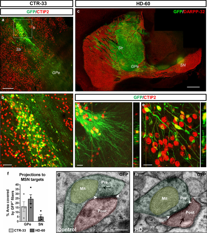
Fig. 2.
Human striatal neurons send axonal projections towards MSN targets and establish synapses within the mouse basal ganglia circuitry. a–e Sagittal sections from CTR-33 and HD-60 chimeric brains at 3 months PST, immuno-labelled for GFP (green), and CTIP2 or DARPP-32 (red). f Histogram representing the percentage of striatopallidal and nigral areas covered by GFP+ human fibres. g, h Ultra-thin immunogold TEM sections showing GFP+ human neurites establishing symmetric inhibitory synapses with host striatal cells. Black arrows point to human-specific gold nanoparticles and white arrowheads delimit the synaptic cleft of symmetric synapses. Str striatum, GPe external globus pallidus, SN substantia nigra, Pre presynaptic terminal, Post postsynaptic terminal, Mit mitochondria. Scale bars 0.5 µm in g; 20 µm in d, e; 50 µm in b; 200 µm in a; 1 mm in c. Data are expressed as mean ± SEM. n = 4 mice

