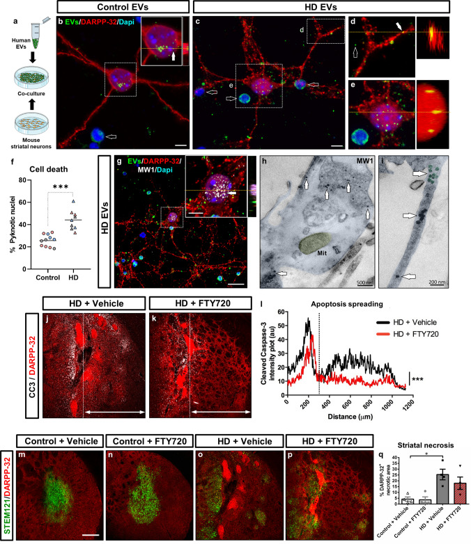
Fig. 7.
HD human neuronal cells secrete extracellular vesicles that propagate toxic soluble mHTT to mouse striatal neurons. a Schematic representation of the co-culture experiment. b–e Co-culture of fluorescently labelled human EVs (green) (filled arrows) isolated from CTR and HD-hNPCs at 16 DIV with DARPP-32+ mouse primary striatal neurons (red), for 24 h at a 2:1 ratio (EV-donor cells: EV-recipient cells). Empty arrows in b, c point to pyknotic nuclei. f Histogram showing the number of pyknotic nuclei in CTR and HD co-cultures. g–i Immunocytochemical and TEM immunogold-labelling for MW1 of mouse striatal neurons co-cultured with human HD EVs, showing the presence of MW1+ monomers/oligomers (white arrows) inside mouse cells. j, k Striatal coronal sections from HD-60 chimeric mice treated with either vehicle or FTY720 at 5 months PST, and immuno-labelled for DARPP-32 (red) and cleaved caspase-3 (white). l Quantification of apoptosis spreading from the bulk of the graft (dotted line in j and k), by analysing cleaved caspase-3 intensity plot profile. m–p Striatal coronal sections from CTR-33 and HD-60 chimeric mice treated with either vehicle or FTY720 at 5 months PST, and immuno-labelled for STEM121 (green) and DARPP-32 (red). q Histogram representing the degree of striatal necrosis in treated vs non-treated chimeric mice. EVs extracellular vesicles, Mit mitochondria. Scale bars 5 µm in b, c; 20 µm in g; 200 µm in m; Data are expressed as mean ± SEM. f n = 11 Control [CTR-33 (n = 5, pink), CTR-2190 (n = 3, grey), GEN-019 (n = 3, blue)] and n = 9 HD [HD-60 (n = 3, pink), HD-2174 (n = 3, grey), GEN-020 (n = 3, blue)] co-cultures; Unpaired t-test, two-tailed, P < 0.0001. l n = 4 mice; Kolmogorov–Smirnov t-test, two-tailed, P < 0.0001. q n = 4 mice; Two-way ANOVA, Tukey’s multiple comparisons test, P = 0.0324 (Control + Vehicle vs HD + Vehicle), P = 0.6367 (HD + Vehicle vs HD + FTY720)

