
Fig. 1.
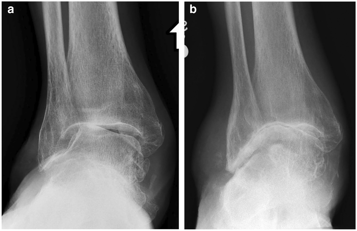
A 65-year-old male patient with history of chronic pes planovalgus deformity, ankle pain, and stiffness had an AP weightbearing radiograph a showing moderate osteoarthritis (OA) of the ankle joint with valgus instability, including lateral predominant joint space narrowing, subchondral sclerosis, and marginal osteophytes. Six years later, the patient presented with worsening pain and decreased range of motion. b Subsequent AP weight-bearing radiograph demonstrates progressive OA with worsening joint space narrowing and marked osseous remodeling resembling a ball-and-socket joint morphology