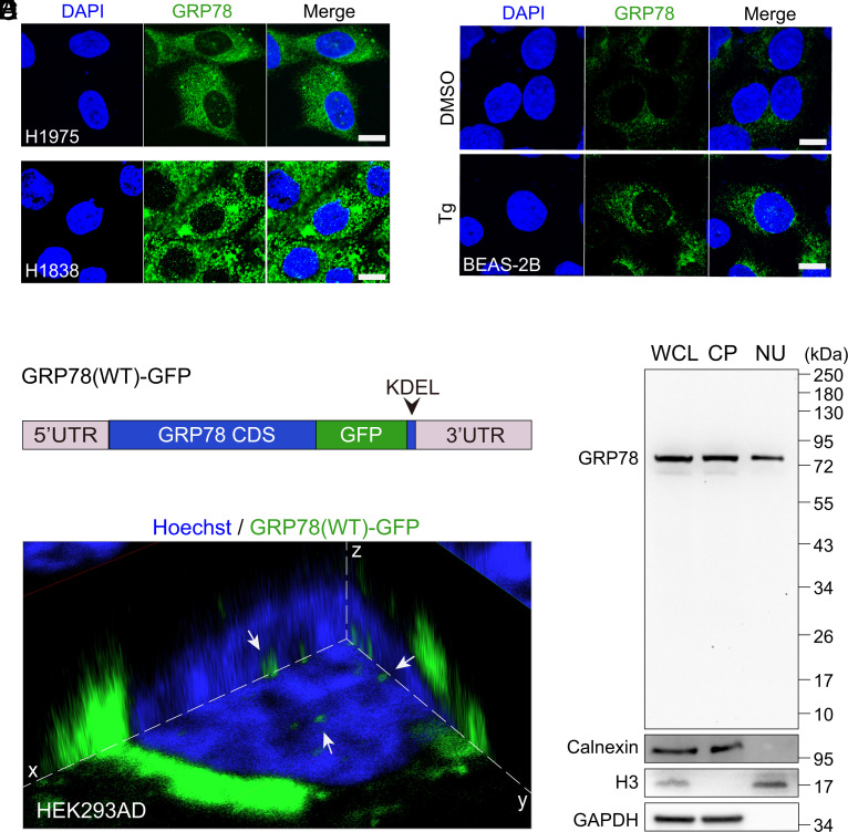
Fig. 3.
GRP78 translocates to the nucleus in human lung cancer cells and ER-stressed normal lung epithelial cells. (A) Representative confocal immunofluorescence images of GRP78 (green) staining in human lung cancer H1975 and H1838 cells. The nuclei were stained by DAPI in blue. (Scale bars, 10 μm.) (B) Representative confocal immunofluorescence images of GRP78 (green) staining in normal human lung epithelial BEAS-2B cells treated with DMSO or thapsigargin (Tg, 100 nM) for 16 h. The nuclei were stained by DAPI in blue. (Scale bars, 10 μm.) (C) Schematic drawing of GRP78(WT)-GFP, which contains the 5′ and 3′ UTRs flanking the full-length, wild-type GRP78 coding sequence (CDS), with the green fluorescent protein (GFP) tag inserted just prior to the KDEL ER retrieval motif. (D) Three-dimensional confocal live cell imaging of HEK293AD cells transfected with the GRP78(WT)-GFP (green) construct for 48 h. The nucleus was stained with Hoechst 33342 (blue). The white arrows indicate GRP78 in the nucleus. (E) Western blot of whole cell lysate (WCL), cytoplasmic (CP), and nuclear (NU) fractions of H1838 cells for GRP78 performed with a polyclonal antibody, with calnexin, Histone H3, and GAPDH serving as ER, nuclear, and cytoplasmic markers, respectively. See also SI Appendix, Fig. S3.

