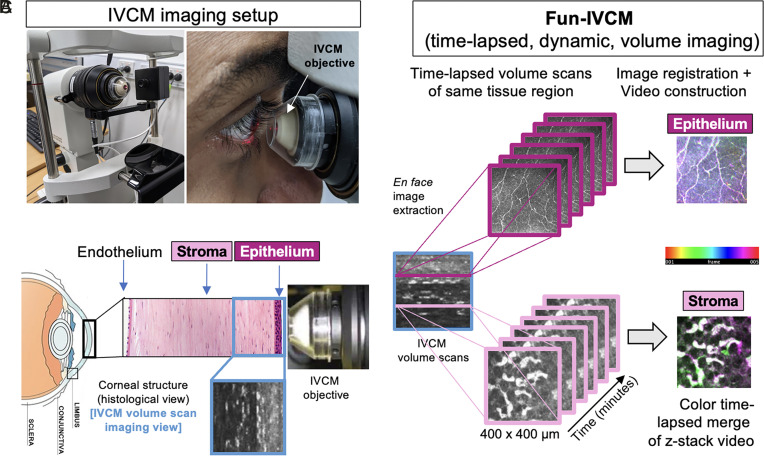
Fig. 1.
Overview of the Fun-IVCM method. (A) Laser-scanning, IVCM setup with the Heidelberg HRT-3 with the Rostock Corneal Module, to acquire images of the human cornea (B), shown diagrammatically in histological cross-section [not to scale]. (C) The Functional IVCM (Fun-IVCM) method involves sequentially capturing high-resolution en face volume (z-stack) images [400(x) × 400(y) × 100(z) µm], spanning the basal layer of the corneal epithelium to the mid-stroma. Repeat volume scans of the same region are acquired every 4 to 7 min for a total of 25 to 40 min. En face images at the same tissue plane are extracted from the volume scans, registered to stationary landmarks, and time-lapsed videos are reconstructed at both the level of the corneal epithelium and anterior stroma.

