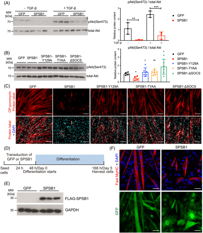
Figure 3.

SPSB1 downregulates TGF‐β signalling by its SPRY and SOCS‐‐box domain and inhibits myogenic differentiation. (A) Five‐day‐differentiated C2C12 myotubes (MT5) transduced with GFP or SPSB1 were treated with TGF‐β (5 ng/mL) or solvent control for 5 min. Lysates were analysed by Western blot analysis with anti‐phospho Akt antibody (Ser473). Total Akt was used as control (left panel). Densitometric analysis (right panel). Data were analysed with two‐way ANOVA followed by Tukey's post‐hoc test. **P < 0.01, ***P < 0.001. (B) C2C12 cells were transduced by control GFP, SPSB1 (WT) or mutants (SPSB1‐Y129A, ‐TYAA or ‐ΔSOCS) retrovirus and differentiated for 5 days. Western blot analysis with anti‐phospho Akt antibody (Ser473) (left panel) and densitometric analysis (right panel). Total Akt was used as control. Data were analysed with one‐way ANOVA followed by Tukey's post‐hoc test. Asterisk (*) indicates significant differences between SPSB1 (wildtype or mutants as indicated) and the GFP control group, *P < 0.05, **P < 0.01, ***P < 0.001; # denotes a significant difference between indicated SPSB1 mutants and the SPSB1 wildtype group, # P < 0.05, ## P < 0.01, ### P < 0.001. (C) O‐Propargyl‐puromycin (OP‐puro) labelling of de novo synthesized polypeptides. Scale bar, 100 μm. (D) Experimental design. (E) Protein lysates from MT5 were analysed by Western blot with anti‐FLAG and anti‐GAPDH antibody. (F) Immunofluorescent staining with anti‐Fast MyHC antibody. Nuclei were stained with DAPI (blue). GFP (green) indicates retrovirally transduced cells. Scale bar, 50 μm.
