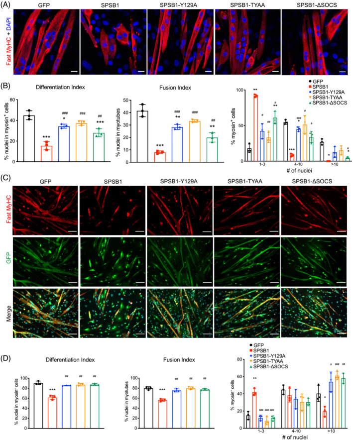
Figure 5.

SPSB1 mediated inhibition of myogenic differentiation depends on its SPRY and SOCS‐‐box domain. (A, B) C2C12 myoblasts were transduced by control GFP, SPSB1 (WT) or mutant (SPSB1‐Y129A, ‐TYAA or ‐ΔSOCS) containing retrovirus and differentiated for 5 days. (A) Immunofluorescent staining with anti‐fast MyHC antibody. Nuclei were stained with DAPI (blue). Scale bar, 20 μm. (B) Differentiation index, Fusion index, and Nuclei distribution in each myosin+ cell were quantified from images in panel (A). (C, D) Primary myoblasts were transduced by control GFP, SPSB1 (WT) or mutant (SPSB1‐Y129A, ‐TYAA or ‐ΔSOCS) containing retrovirus and differentiated for 5 days. (C) Immunofluorescent staining with anti‐fast MyHC antibody (red). GFP (green) indicates retrovirally transduced cells. Scale bar, 100 μm. (D) Differentiation index, Fusion index, and Nuclei distribution in each myosin+ cell were quantified from images in panel (C). Data in panels (B and C; Differentiation and Fusion index), were analysed with two‐tailed Student's t‐test; data in panels (B) and (H) (Nuclei distribution in myosin+ cells) were analysed with two‐way ANOVA followed by Tukey's post‐hoc test; asterisk (*) indicates significant differences between SPSB1 (wildtype or mutants as indicated) and the GFP control group, *P < 0.05, **P < 0.01, ***P < 0.001; denotes a significant difference between indicated SPSB1 mutants and the SPSB1 wildtype group, # P < 0.05, ## P < 0.01, ### P < 0.001. N = 3 biologically independent experiments; data are presented as mean ± standard deviation.
