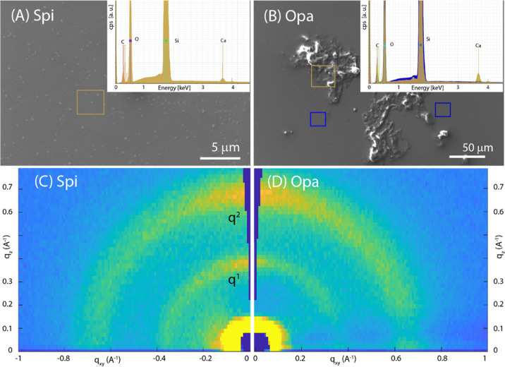
Figure 3.
(A,B) SEM images of SOM spin-coated on a silicon wafer from S. pistillata (Spi) and O. patagonica (Opa), respectively. In the inset the EDX spectra recorded from the square regions drawn on the SEM images are reported. (C,D) 2D-GIWAXS images from Spi and Opa, respectively, deposited on the substrate. q1 and q2 indicate the diffraction peaks analyzed in Table 1.

