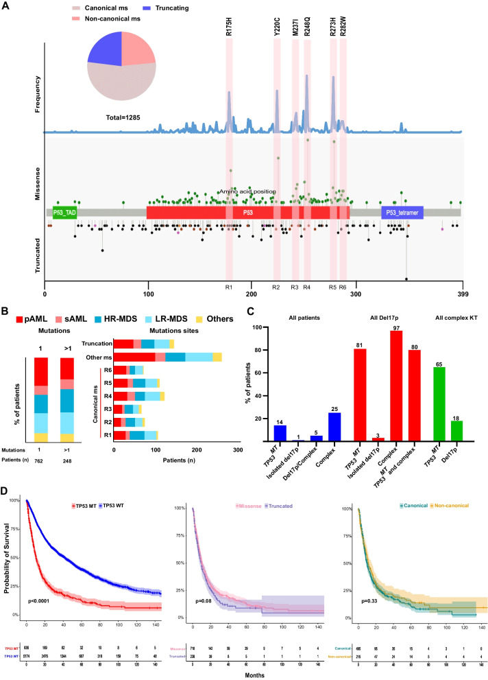
Fig. 1.
Distribution of TP53 mutations with hotspot locations and chromosome 17 deletion. A Schematic drawing of TP53 gene showing the location of mutations, the type of mutations, and canonical sites. Frequency of mutations in all cohort is shown in the upper part. Missense and truncated mutations are indicated in the upper and lower part of the gene structure, respectively. B Bar graphs showing the frequencies of single (1) and multiple TP53 mutations (> 1) and canonical missense locations in each disease subtype. C Percentage of patients with cytogenetics abnormalities in relation to the TP53 mutational status. D Kaplan–Meier survival curves of patients with TP53 mutations vs. wild type, missense vs. truncated mutations, and canonical vs. non-canonical missense mutations

