Fig. 3.
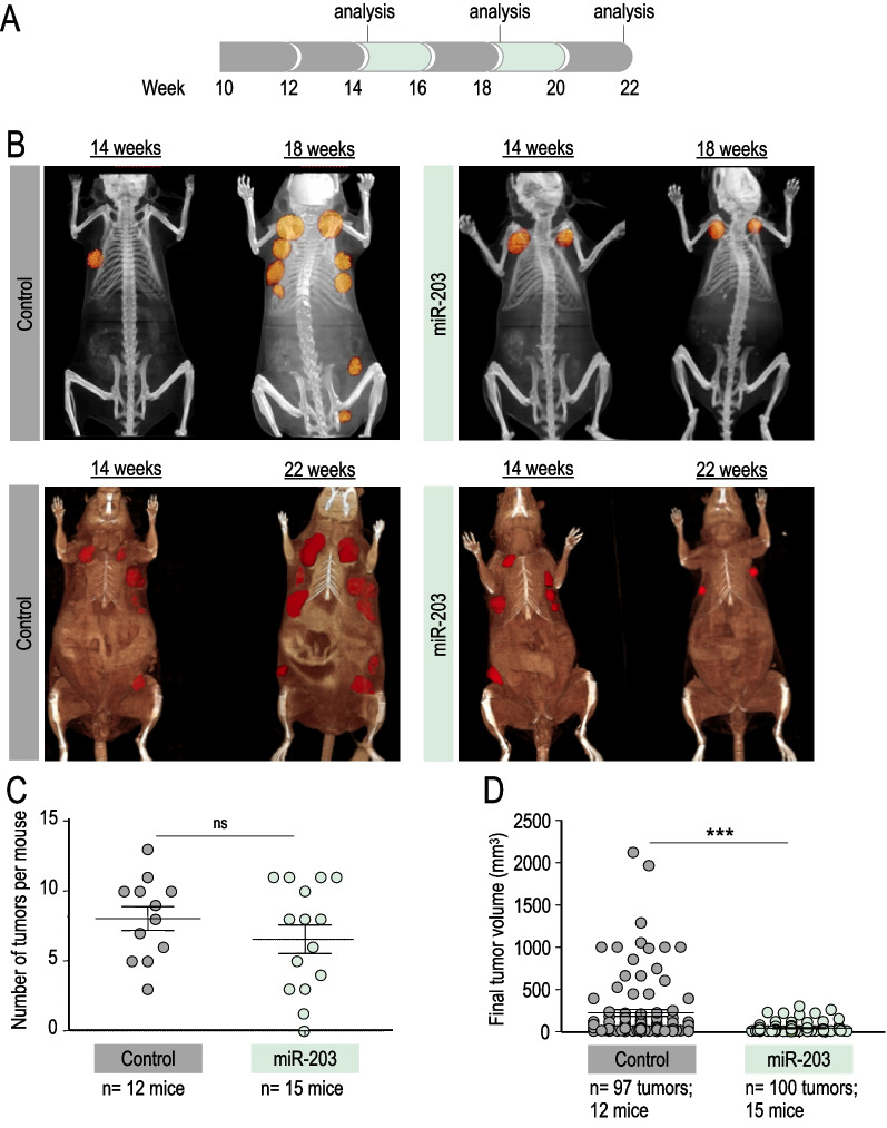
In vivo effects of miR-203 treatment on PyMT mice, started at tumor CT detection and administered every two weeks. A Schematic of the Dox treatment (in green) schedule in vivo, on miR-203 wild-type or miR-203 knock-in; PyMT mice, starting when tumors are found by micro-CT imaging (around week 14) to the endpoint, on alternating weeks. B Representative micro-CT images of mice subjected to the Dox treatment (in the figures, “control” indicates miR-203 wild-type; “miR-203” indicates knock-in mice) at tumor detection by micro-CT (14 weeks), four weeks later (18 weeks) and at the endpoint (22 weeks). C Number of tumors per mouse at the endpoint, in control and miR-203-treated mice. D, Final tumor volume of control and miR-203-treated mice. In C, D, data are represented as mean ± s.d. (Number of mice and total number of tumors per group are indicated in the figure.) ***p < 0.001; n.s. not statistically different (Student’s t test)

