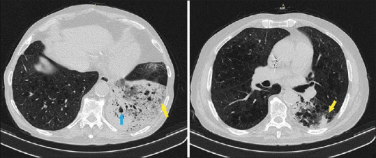
Figure 3.

In the chest Computed Tomography (CT), it is reported emphysematous lesions (light blue arrow) and increased consolidation with air bronchogram and multiple emphysema cysts in the left lower lobe. The foci in the lower left lobe was increased (yellow arrows)
