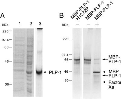
FIG. 2.
Overexpression and purification of recombinant PLP-1. Proteins were purified as described in Materials and Methods, analyzed by SDS–10% (A) or 4 to 15% (B) polyacrylamide gel electrophoresis, and stained with Coomassie blue. (A) Expression of PLP-1 from pET-PLP-1. Lane 1, supernatant after sonication and centrifugation; lane 2, pellet after sonication and centrifugation; lane 3, refolded PLP-1. PLP-1 is indicated by the arrow. (B) Purified MBP–PLP-1 H1272P and MBP–PLP-1 with (−) and without (+) factor Xa cleavage. MBP–PLP-1 (or MBP–PLP-1 H1272P), MBP, PLP-1, and factor Xa are indicated by arrows. The molecular masses of marker proteins are indicated to the left of each panel.

