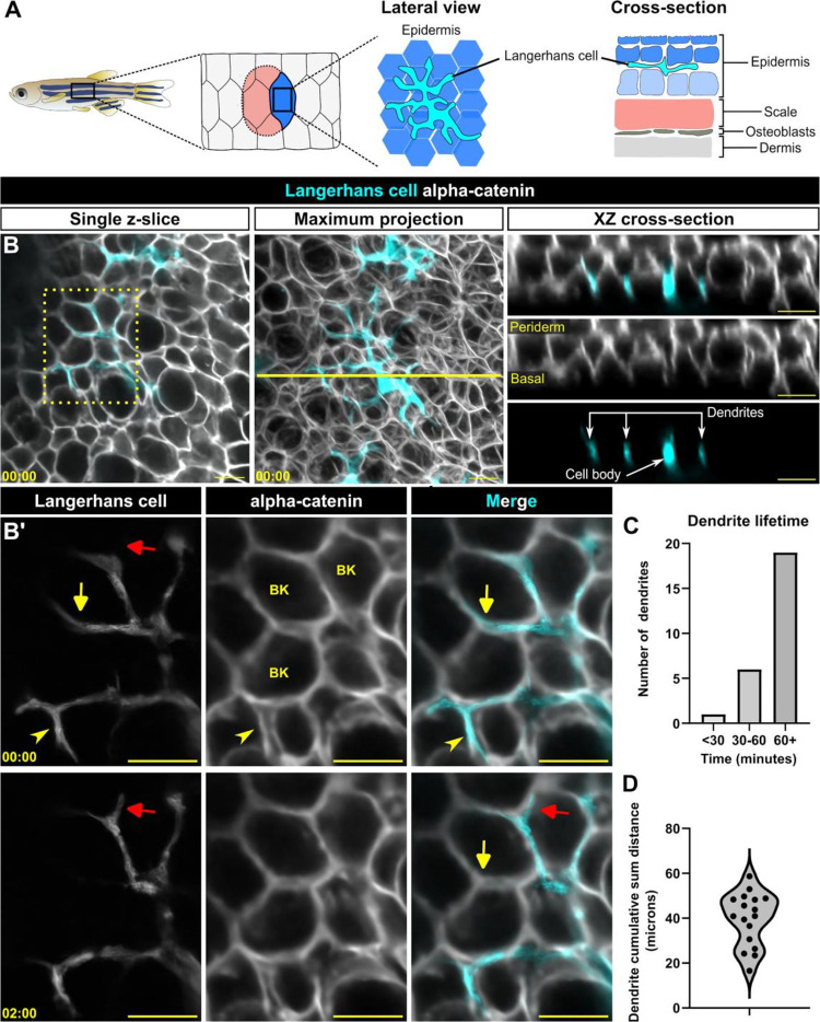
Figure 1. Langerhans cell dendrites survey the epidermis in the absence of stimulus.
A. Schematic of adult zebrafish trunk skin, showing lateral and cross-section views of the epidermis. B. Single z-slice (left), maximum intensity projection (middle) and YZ projection along yellow line in middle panel (right) images of Tg(mpeg1:NTR-EYFP)-positive cells and Gt(ctnna1-Citrine) labeled epidermal cell membranes, illustrating the complexity of skin epidermis. B’. Inset from dashed box in B (single z-slice), illustrating Langerhans cell dendrite retraction. See also Supplemental Video 1. Yellow arrow denotes dendrite retraction over time, red arrow denotes dendrite extension over time, arrowhead denotes a dendrite occupying space between keratinocytes. BK, basal keratinocyte. C. Quantification of dendrite lifetime, n = 26 dendrites tracked from 7 cells. D. Violin plot of cumulative sum distance traveled by dendrites over a 10 minute window, n = 17 dendrites tracked from 7 cells. Timestamps in (B, B’) denote mm:ss. Scale bars in (B, B’) denote 10 microns.

