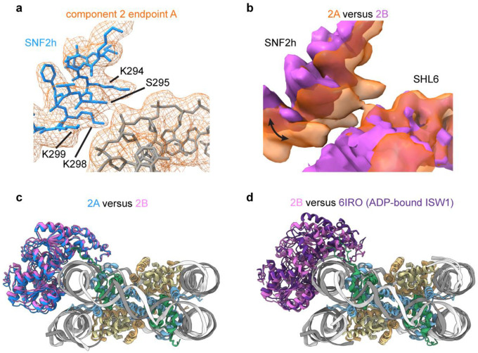
Extended Data Fig. 9. SNF2h rocking to and from SHL6.
a) Interaction interface between SNF2h and nucleosomal DNA at SHL6 observed in endpoint structure 2A from 3DVA of the single-bound SNF2h-nucleosome particles. Clear contacts between DNA and residues K294, S295, K298, and K299 of SNF2h are observed.
b) Overlay of coulomb potential maps for endpoint 2A and 2B illustrating movement of SNF2h to and from SHL6.
c) Overlay of the models for single-bound SNF2h-nucleosome complex endpoint structures 2A and 2B. The entire SNF2h ATPase domain rocks upwards in structure 2B (pink) versus structure 2A (blue).
d) Overlay of the models for single-bound SNF2h-nucleosome complex endpoint structure 2B and the previously determined ADP-bound ISW1-nucleosome structure (PDB 6IRO). While the bottom ATPase lobe (lobe 1) appears similarly positioned and dissociated from SHL6, the top ATPase lobe (lobe 2) dramatically shifts upwards in the ADP-bound structure.

