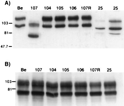
FIG. 2.
Expression of gE proteins. PK15 cells were infected at an MOI of 10 with either PRV Be (wild type), PRV 107 (Am457), PRV 104 (Y478S), PRV 105 (Y478S + Y517S), PRV 106 (Y517S), PRV 107R (revertant PRV 107), or PRV 25 (frameshift) for 16 h prior to preparation of cell lysates. Western blot analysis was performed with either rabbit polyclonal antiserum to gE (A) or goat polyclonal antiserum to gC (B). In lanes 1 to 7 of panel A, 10 μg of total protein was loaded in each lane; 60 μg of total protein was loaded in lane 8. Immature wild-type gE protein has a molecular mass of approximately 93 kDa and a mature form of approximately 110 kDa. Cells infected with PRV 107 contain immature and mature forms of approximately 70 and 93 kDa, respectively. The gE protein produced by PRV 25 has an immature form of 80 kDa and a mature form of 101 kDa. In panel B, 10 μg of total protein was analyzed for each sample. Positions of apparent molecular mass markers are shown on the left in kilodaltons.

