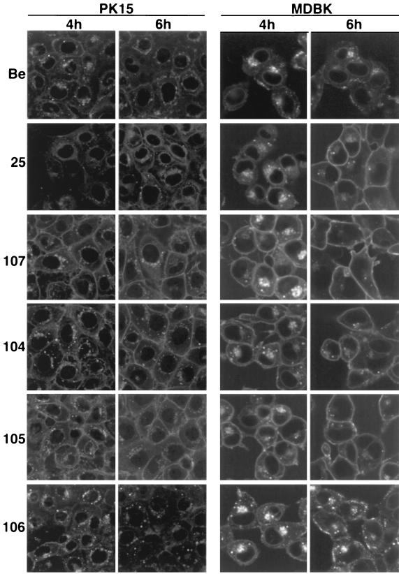
FIG. 3.
Steady-state distribution of the gE-gI complex. PK15 and MDBK cells were infected at an MOI of 10 for the times indicated with either PRV Be (wild type), PRV 25 (frameshift), PRV 107 (Am457), PRV 104 (Y478S), PRV 105 (Y478S + Y517S), or PRV 106 (Y517S). The cells were fixed, permeabilized, and reacted with a MAb that specifically recognized gE when it was complexed with gI (MAb 1/14). An Alexa-568-conjugated secondary antibody was used to visualize bound MAb. Confocal sections were taken through the centers of the cells.

