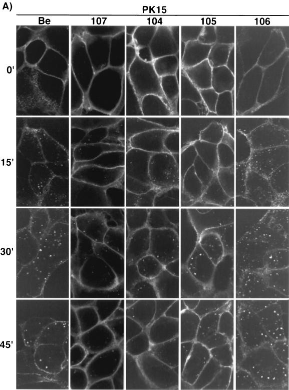
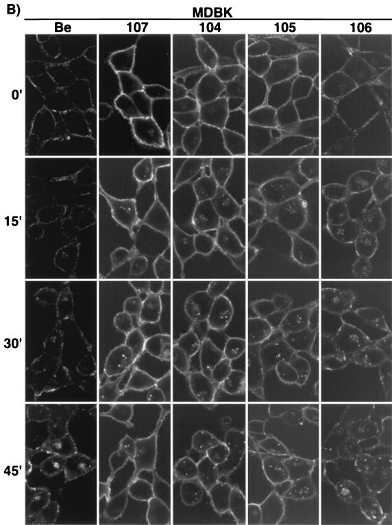
FIG. 4.
Endocytosis of the gE mutant-gI complex. PK15 (A) and MDBK (B) cells were infected at an MOI of 10 with either PRV Be (wild type), PRV 107 (Am457), PRV 104 (Y478S), PRV 105 (Y478S + Y517S), or PRV 106 (Y517S) for 4 h prior to an indirect immunofluorescence endocytosis assay as described in Materials and Methods. Briefly, the cells were incubated at 4°C with MAb 1/14, which specifically recognized gE when it was complexed with gI prior to a shift of the cells to 37°C for the indicated times to allow internalization of the protein complex. The cells were then fixed, permeabilized, and reacted with an Alexa-568-conjugated secondary antibody to visualize the bound primary antibody. Confocal sections were taken through the centers of the cells.


