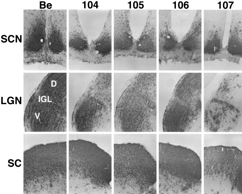
FIG. 6.
Localization of viral antigen in brain sections. The brains from animals taken from infections described in the legend to Fig. 7 were removed and analyzed for viral antigen with a polyvalent rabbit antiserum generated against whole virus particles (Rb133). Serial sections (35 μm) through the coronal plane were cut, processed and mounted on slides. Representative sections containing the SCN, LGN including the dorsal (D) and ventral (V) aspects as well as the IGL, and the SC are pictured.

