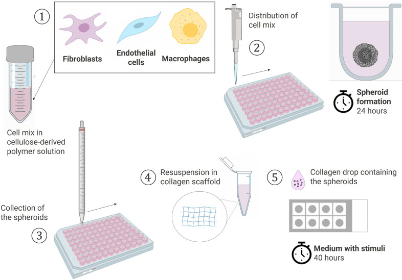
Figure 1.
Schematic workflow of the spheroid formation and the sprouting assay in a collagen-based 3D scaffold. (1) Cells pooled together in 20% Methocell solution (2) Spheroid formation: distribution of 150 µl cell mix/well into a 96 U-well suspension plate and incubation at 37°C for 24h (3) Collection of the spheroids with a 10 ml pipet (4) Spheroids resuspended in a 1.5 mg/ml collagen solution (5) Sprouting assay: 20 µl of collagen solution containing the spheroids placed dropwise using the sandwich method in a 8-chamber slide and cultured in the suitable media containing stimuli at 37°C for 40h. Created with BioRender.com.

