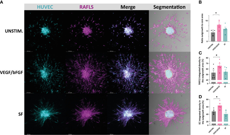
Figure 2.
Application of a machine learning quantitative analysis (QuPath) in the pre-established spheroid-based model of RA synovial angiogenesis to measure spheroid outgrowth and morphological changes. Spheroids were either left unstimulated (UNSTIM.), stimulated with VEGF/bFGF (10 ng/ml) or SF (20%). Representative confocal Z-stack projection pictures (10X) of the 3D model containing 3.75x10⁴ RAFLS (magenta) and 7.5x10⁴ EC (cyan), including the specific fluorescence signal for each cell type and the output segmentation of the outgrowth area (pink) versus the core area (blue) using trained pixel classifiers in QuPath (A). Ratio of the spheroid outgrowth area to core area (n=5) (B). Percentage of the total integrated density of RAFLS (C) and ECs (D) calculated in the outgrowth area (n=5). Integrated density is the product of mean fluorescence intensity and area. Statistical significance was determined by RM one-way ANOVA (*=p<0.05).

