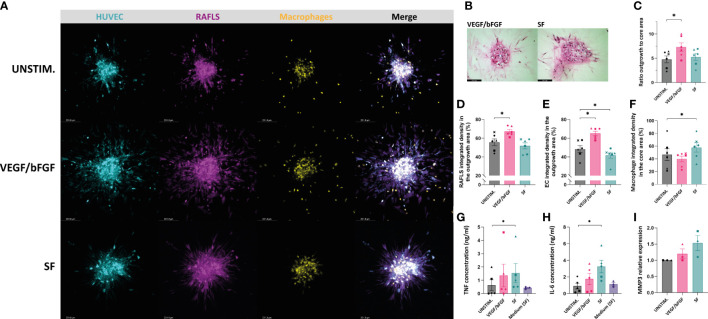
Figure 4.
VEGF/bFGF significantly induces spheroid outgrowth in the new 3D model containing macrophages, whereas RASF enhances macrophage containment and compaction. Spheroids were left either unstimulated (UNSTIM.), stimulated with VEGF/bFGF (10 ng/ml) or SF (20%). Representative confocal Z-stack projection pictures of the 3D model containing 3.75x10⁴ RAFLS (magenta), 7.5x10⁴ EC (cyan) and 3x10⁴ macrophages (yellow) (A). Representative pictures of H&E staining in paraffin-embedded spheroid sections for the stimulated condition (5 µm) (B). Ratio of the spheroid outgrowth area to core area (n=6) (C). Percentage of the total integrated density of RAFLS (D) and ECs (E) calculated in the outgrowth area (n=6). Percentage of the total integrated density of macrophages calculated in the core area (n=6) (F). Concentrations of TNF and IL-6 proteins in the spheroid supernatants as detected by ELISA (n=5) (G, H). Relative mRNA expression of MMP3 expressed in fold-change value compared to the unstimulated condition (n=3). The relative expression was normalized to both GAPDH and RPLP0 reference genes using ΔΔCT method (average of the two fold-change values) (I). Statistical significance was determined by RM one-way ANOVA for the analysis of outgrowth ratio and integrated density of RAFLS/EC and by ratio paired t-test for the integrated density of macrophages. A Friedman test was applied for TNF and IL-6 protein concentrations (*=p<0.05).

