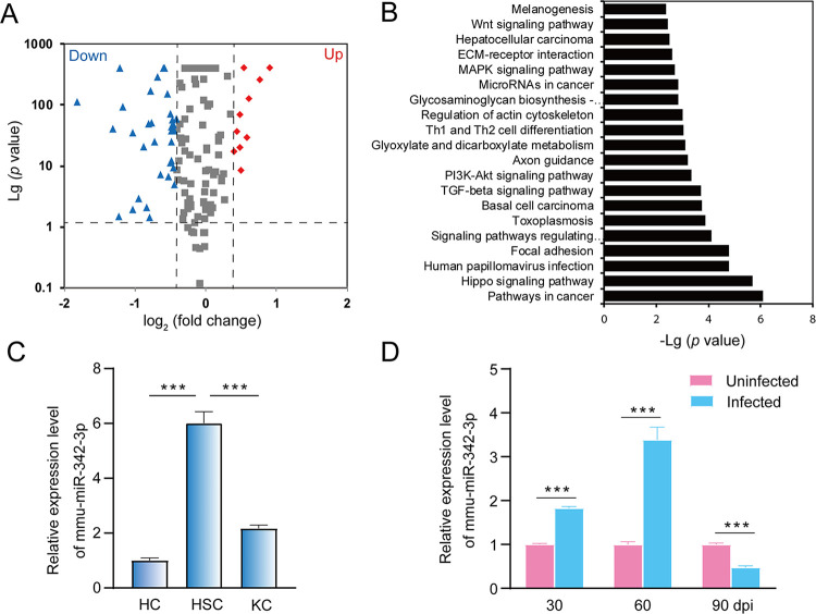
Fig 3. Dysregulation of mmu-miR-342-3p in liver HSCs during E. multilocularis infection.
(A) A volcano plot of the differentially expressed miRNAs between two liver HSCs from E. multilocularis-infected mice at 60 and 90-day post infection (dpi), where the red and green, indicated significantly upregulated miRNAs (Up) and downregulated miRNAs (Down), respectively. (B) KEGG pathway enrichment analysis of target genes. (C) The expression of mmu-miR-342-3p in hepatocyte cells (HCs), Kupffer cells (KCs), and hepatic stellate cells (HSCs). (D) The expression of mmu-miR-342-3p in liver HSCs from E. multilocularis-infected mice at 30-, 60-, and 90-day post-infection (dpi). The uninfected mice at each sampling time point were used as controls. Data for final statistical analysis were taken from 3 independent experiments. ***p < 0.001.

