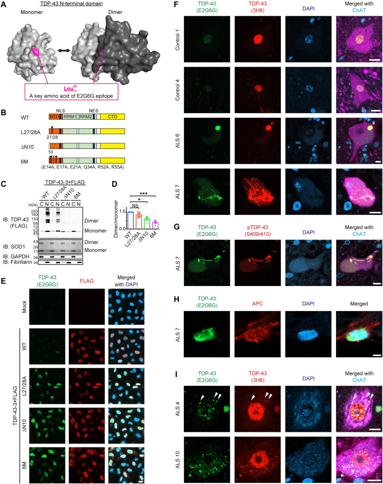
Fig. 2. NDD-TDP-43 constitutes pathological inclusion bodies in ALS.
(A) Three-dimensional models of the monomeric and dimeric NTD of TDP-43 as predicted by AlphaFold2. The critical amino acid of the anti–TDP-43 monoclonal antibody (E2G6G) epitope, Leu41, is indicated. (B) Schematic illustration of the TDP-43WT and three N-terminal dimerization–deficient mutants (NDD mutants). NLS, nuclear localization signal; NES, nuclear export signal. (C) Representative immunoblots of DSG–cross-linked subcellular fractions obtained from Neuro2a cells transiently expressing TDP-43-3×FLAG WT or NDD mutants. Monomeric, dimeric, and multimeric TDP-43 in the cytoplasmic (C) and nuclear (N) fractions were detected using an anti-FLAG antibody. SOD1, GAPDH (glyceraldehyde-3-phosphate dehydrogenase), and fibrillarin were used as markers for equal cross-linking, cytoplasmic fractions, and nuclear fractions, respectively. (D) Quantification of the dimer/monomer ratio of TDP-43 (relative to TDP-43WT) in (C). n = 4, biologically independent experiments. Data are expressed as means ± SEM. *P < 0.05 and ***P < 0.001 [analysis of variance (ANOVA) with Tukey’s test]. (E) Representative images of HeLa cells transiently expressing TDP-43 WT or NDD mutants immunostained with E2G6G and anti-FLAG antibody. DAPI, 4′,6-diamidino-2-phenylindole. (F) Representative images of spinal motor neurons from two controls and four patients with sporadic ALS (sALS), with similar results within the groups, immunostained with E2G6G, anti–panTDP-43 (3H8), and anti–choline acetyltransferase (ChAT). (G) Representative images of a spinal motor neuron from a patient with sALS immunostained with E2G6G, anti–pTDP-43 (Ser409/Ser410), and anti-ChAT. (H) Representative images of an oligodendrocyte in the spinal cord from a patient with sALS immunostained with E2G6G and anti-APC. (I) Representative images of spinal motor neurons from a patient with sALS with remaining nuclear TDP-43, immunostained with E2G6G, 3H8, and anti-ChAT. Arrowheads indicate E2G6G-positive cytoplasmic granules. Scale bars, 20 μm (E), 10 μm (F, G, and I), and 2 μm (H).

