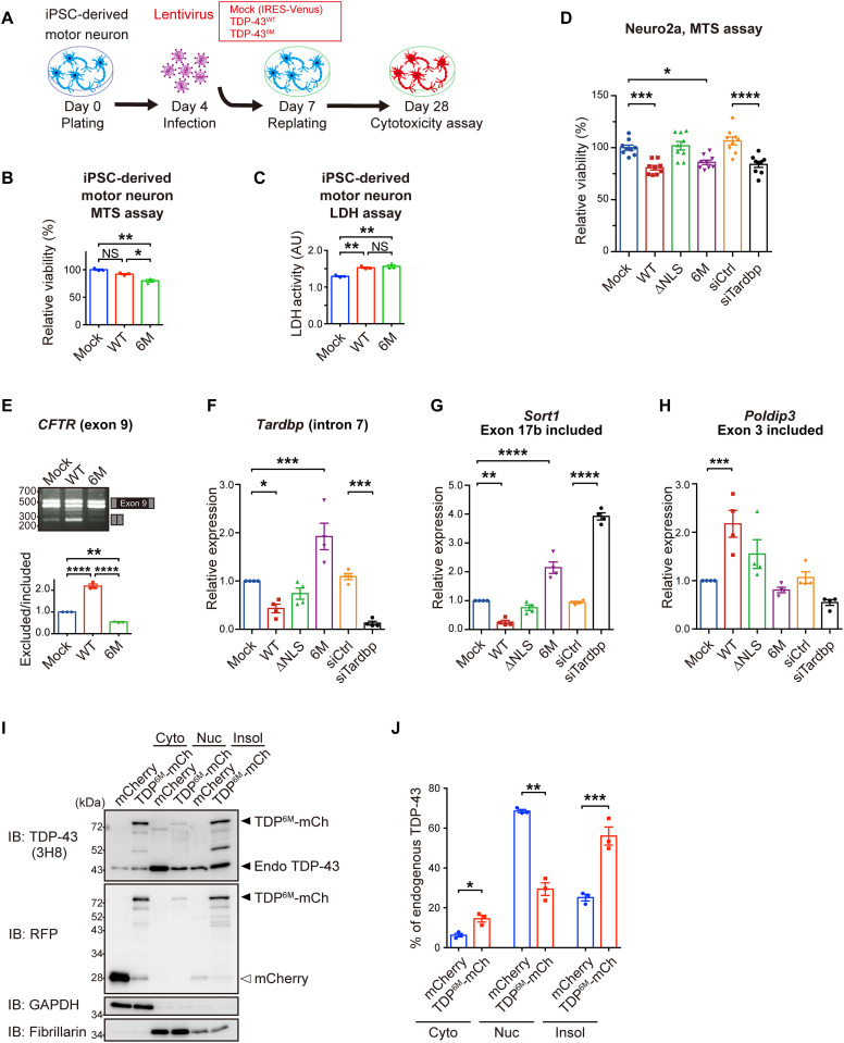
Fig. 6. NDD-TDP-43 mutant impairs endogenous TDP-43 function by sequestrating the endogenous TDP-43 into aggregates.
(A to C) Cell viability and cytotoxicity of iPSC-derived motor neurons infected with lentivirus expressing TDP-43WT or TDP-436M. Experimental protocol (A), MTS assay (B), and LDH assay (C) (relative to mock-transfected cells). (D) Relative viability of Neuro2a cells transiently transfected with TDP-43, control siRNAs, or siRNA targeting TDP-43 (relative to mock-transfected cells) was determined by MTS assay. Triplicate samples were analyzed in three biologically independent experiments. (E) Exon skipping assay for CFTR exon 9. Neuro2a cells were transiently cotransfected with TDP-43 and CFTR minigene reporter plasmids. Splicing of CFTR exon 9 was assessed by RT-PCR (top). Quantification of the exclusion/inclusion ratio of CFTR exon 9 (relative to mock-transfected cells) was performed from the band intensities (bottom). n = 3, biologically independent experiments. (F to H) Quantification of RNA levels of Tardbp intron 7 (F), Sort1 (G), and Poldip3 (H) with indicated exon inclusion. Neuro2a cells were transiently transfected with TDP-43, control siRNAs, or siRNAs targeting TDP-43, and mRNA levels (relative to mock-transfected cells) were analyzed by quantitative RT-PCR. n = 4 biologically independent experiments. (I) Representative immunoblots showing endogenous TDP-43 and mCherry levels in cytoplasmic (Cyto), nuclear (Nuc), and 1% Triton X-100 insoluble fractions (Insol) of Neuro2a cells transiently expressing mCherry or TDP-436M-mCherry. GAPDH and fibrillarin were used as the cytoplasmic and nuclear markers, respectively. (J) Quantification of the percentage of endogenous TDP-43 in each fraction to the total endogenous TDP-43 in (I). n = 3, biologically independent experiments. Data are expressed as means ± SEM. *P < 0.05, **P < 0.01, ***P < 0.001, and ****P < 0.0001 [ANOVA with Tukey’s test in (B) to (H) and unpaired two-sided t test in (J)].

