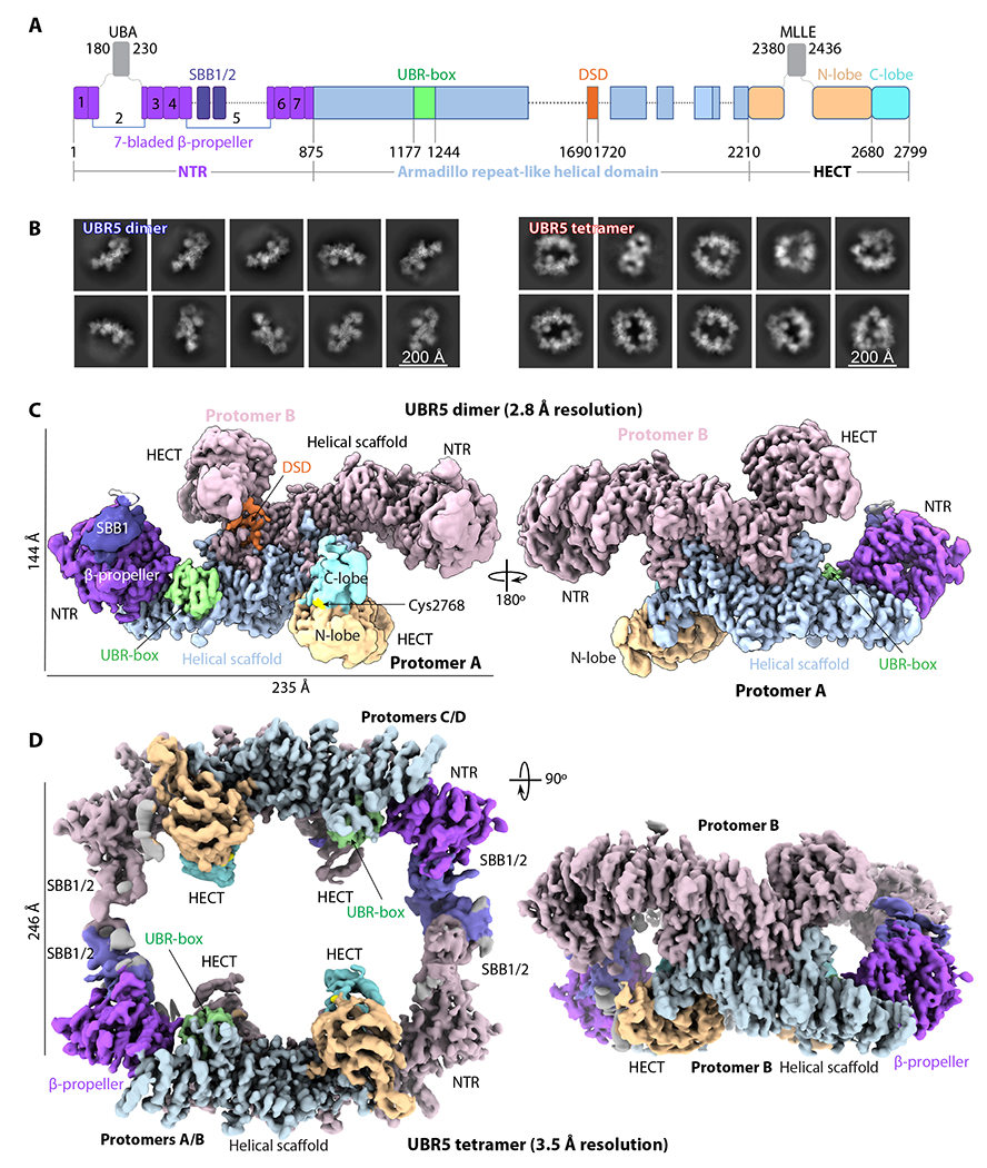
Figure 1. Cryo-EM of the UBR5 dimer and tetramer.

(A) Domain architecture of the human E3 ligase UBR5. Unresolved regions in the EM map are shown as dash lines. The disordered UBA and MLLE domains are shown as gray squares. (B) Selected 2D class averages of the dimer (left) and tetramer (right). (C-D) Cryo-EM 3D maps of the dimer (C) and tetramer (D). Domains are colored as in (A).
