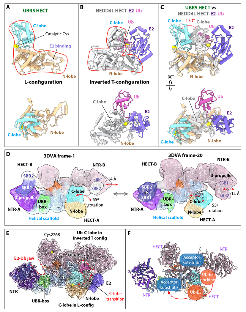
Figure 5. UBR5 undergoes large conformational changes around the E2–Ub jaw.

(A) UBR5 HECT domain in the L-conformation in two orthogonal views. (B) The NEDD4L-HECT-E2-Ub structure (PDB ID 3JWO) in the same view. (C) Superimposition of the UBR5-HECT (this study) and NEDD4L-HECT-E2-Ub (PDB ID 3JWO) structures. The UBR5 HECT N-lobe is poised to bind E2, but the C-lobe needs to rotate 130° to reach the C-lobe position of the NEDD4L-HECT for transthiolation reaction. (D) The two most distinct conformations of the UBR5 dimer as determined by 3DVA, showing a 14 Å lateral movement of the NTR and a 55° rotation of the HECT. SBB2 above SBB1 was observed in this lower resolution variability analysis, but missing in the 2.8 Å 3D map, indicating its high mobility in the dimer. (E) Ub-E2 docked in the right intermolecular jaw of the UBR5 dimer. The distance between the C-lobe and UBR-box is 62 Å. (F) Possible substrate ubiquitylation pathway. The curved red arrow indicates that E3 Ub transthiolation reaction occurs in the intermolecular jaw. The dashed red arrow indicates the Ub transfer route for substrate ubiquitylation.
