Figure 1.

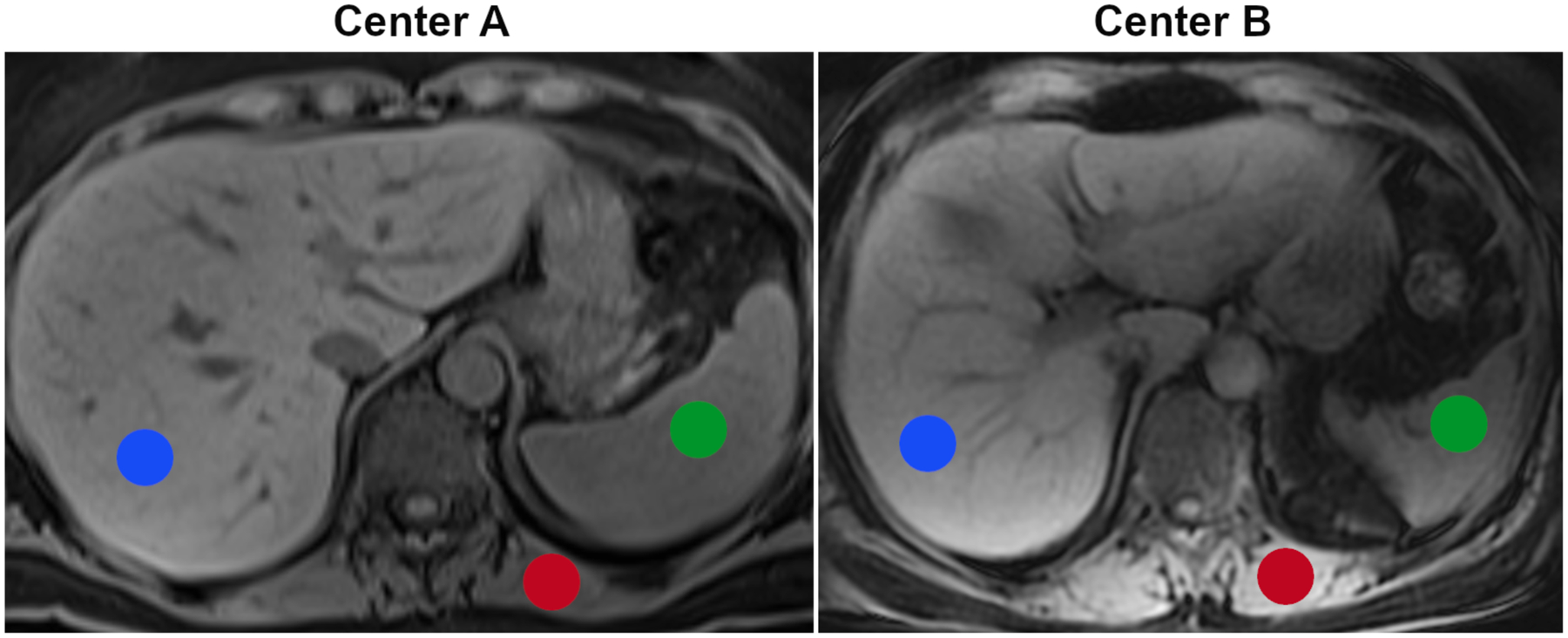
Examples for T1-weighted Dixon MR images from the two centers and volumes of interest for the three tissues of interest (liver, red; spleen, green; and paraspinal muscle, red).

Examples for T1-weighted Dixon MR images from the two centers and volumes of interest for the three tissues of interest (liver, red; spleen, green; and paraspinal muscle, red).