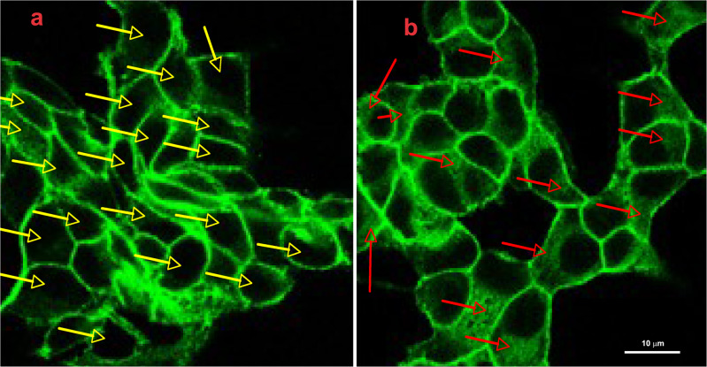
Fig. 4.
Following GlcN treatment cytosolic β-catenin increased. INS-1E cells were grown on glass coverslips for 48 h, then were vehicle-treated (a) or treated with 7.5 mM GlcN for 24 h (b). Cells were stained with anti-β-catenin antibodies. Yellow arrows in the control cells indicate cells without intracellular β-catenin, red arrows in treated cells indicate cells with intracellular β-catenin. Following GlcN treatment there was the appearance of intracellular β-catenin. n = 2

