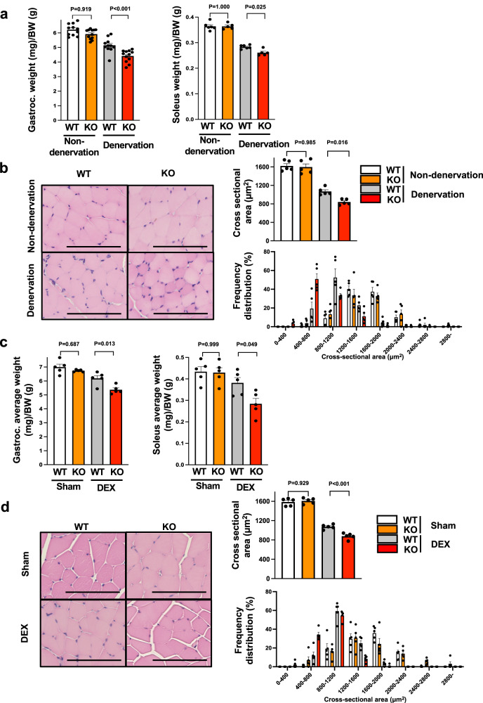
Fig. 2. Myonectin deficiency exacerbates muscle atrophy induced by sciatic nerve denervation or steroid administration.
a, b WT and myonectin-KO mice at the age of 8–10 weeks were subjected to sciatic denervation-induced muscle atrophy operation. At 5 days after sciatic nerve denervation, the denervated and non-denervated (contralateral) muscles were used for analyses. a Gastrocnemius (n = 11 mice per group) or soleus (N = 6 in non-denervated WT, non-denervated KO and denervated WT mice, N = 5 in denervated KO mice) muscle weight/body weight ratio is shown. b Left panels show representative cross sectional images of gastrocnemius muscle fibers. Scale bars show 100 μm. Right panels show quantitative analyses of mean cross sectional area (CSA) and CSA distribution of gastrocnemius muscle. N = 5 in each group. c, d At 14 days after continuous administration of dexamethasone (DEX) or vehicle (Sham), WT and myonectin-KO mice were sacrificed for analysis. c The ratio of gastrocnemius or soleus muscle weight to body weight is shown. N = 5 in each group. d Left panels show representative cross sectional images of gastrocnemius muscle fibers. Scale bars show 100 μm. Right panels show quantitative analyses of mean CSA and CSA distribution of gastrocnemius muscle. N = 5 in each group. Data are presented as means ± SEM. One-way ANOVA with a post-hoc analysis for a–d was performed. Gastroc. gastrocnemius muscle, BW body weight, DEX dexamethasone.

