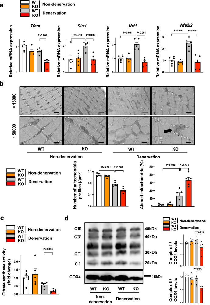
Fig. 4. Myonectin deficiency leads to exacerbated mitochondrial dysfunction in denervated muscle.
a–d WT and myonectin-KO mice at the age of 8–10 weeks were subjected to sciatic denervation-induced muscle atrophy operation. At 5 days after sciatic nerve denervation, the denervated and non-denervated gastrocnemius muscles were used for analyses. a The mRNA levels of genes related to mitochondrial biogenesis were evaluated by quantitative real time PCR method. N = 6 in non-denervated and denervated WT mice. N = 5 in non-denervated and denervated KO mice. b Upper panels show representative electron micrographs of gastrocnemius muscles. Arrow shows altered mitochondria. Altered mitochondria is defined as a mitochondria which has partial or complete separation of the outer and inner membranes and swelling. Lower panels show quantification of intra-myofibrillar mitochondria number and altered mitochondria percentage. N = 5 in each group. Scale bars show 1μm in X15,000 and 400 nm in X50,000. c Relative citrate synthase activity in isolated mitochondrial fraction from denervated gastrocnemius muscles was measured. N = 6 in non-denervated and denervated WT mice. N = 5 in non-denervated and denervated KO mice. d Left panel shows representative Western blot analyses of oxidative phosphorylation complexes. Right panels show quantitative analyses of the complex I and complex II signals. N = 5 in each group. Data are presented as means ± SEM. One-way ANOVA (a, b) with a post-hoc analysis and two-tailed unpaired Student’s t-test (c, d) were performed.

