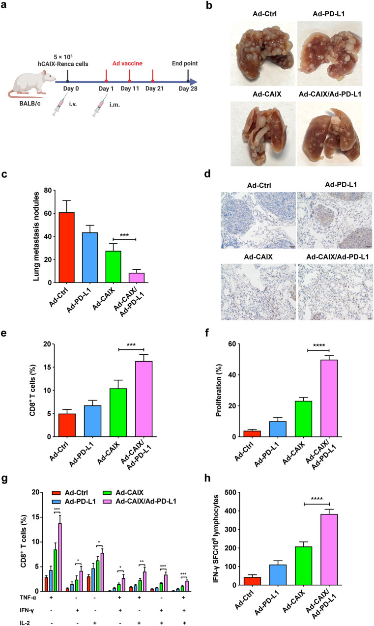
Fig. 5. The therapeutic effects of Ad-CAIX/Ad-PD-L1 in the hCAIX-Renca lung metastasis model.
a Schematic diagram illustrating the establishment of lung metastasis model and the experimental design. b Representative images of metastatic nodules on lung surface of mice each group. c Quantitative statistical analysis of the number of nodules on the lung surface. d Immunohistochemistry was used to detect CD8+ T cell infiltration in the lungs of mice in each group (scale bar, 50 μm). e Percentages of CD8+ T cells in infiltrating immune cells from the lung tumor tissues of mice in each group. f The proliferation of CD8+ T cells was detected by the EdU assay. g The percentages of CD8+ T cells expressing TNF-α, IFN-γ, or IL-2 in stimulated splenocytes. h IFN-γ- secreting T lymphocyte cells were detected by ELISPOT assay. Data were from one representative experiment of three performed and presented as mean ± SD. The different significance was set at **p < 0.01 and ***p < 0.001. Multiple groups of comparison data were analyzed by one-way ANOVA.

