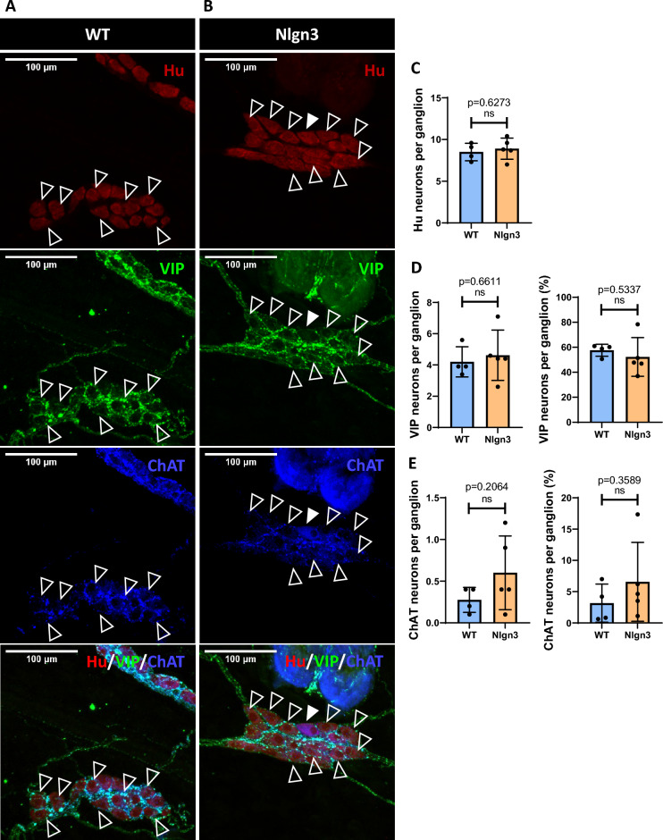
Figure 6.
Hu, VIP, and ChAT immunostaining in the submucosal plexus of WT (A) and Nlgn3R451C (B) mice. VIP (clear arrowheads) and ChAT (filled arrowhead) immunolabelled enteric neurons in the cecal submucosal plexus are pictured. (C) No significant difference was observed in the number of Hu immunolabelled neurons per ganglion. (D) The number of VIP immunostained neurons and the percentage of VIP immunostained neurons per ganglion was not significantly different. (E) No significant difference was observed in the number of ChAT immunolabelled neurons and the percentage of ChAT neurons per ganglion. Statistical comparisons were conducted using Student’s unpaired t-test and individual data and mean ± SD were plotted for n = 11 mice in each group. ns = p > 0.05. WT = wildtype; Nlgn3 = Nlgn3R451C.

