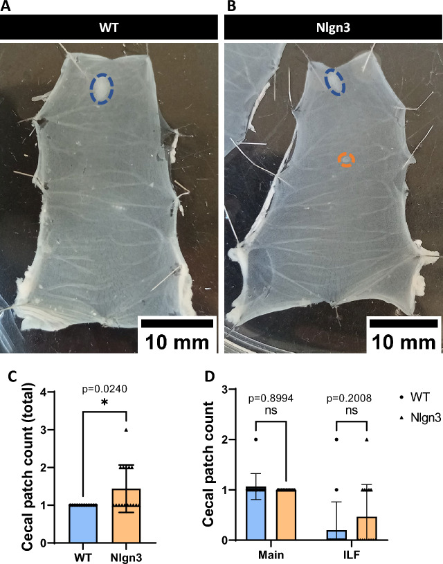
Figure 7.
Multiple cecal patches are present in Nlgn3R451C mice.Representative images of pinned-out cecum from WT (A) and Nlgn3 (B) mice. Blue dashes outline the main cecal patch, whereas orange dashes outline the isolated lymphoid follicles (ILF) typically found along the body of the cecum. (C) Total cecal patch numbers are significantly higher in Nlgn3 cecum compared with WT littermates. 3 outliers were removed in the WT group according to the ROUT Q = 1% method. (D) When total cecal patch count is split into main and ILF cecal patches, the difference is not significant despite an evident, qualitative trend for an increase in Nlgn3 ILF. Statistical comparisons were conducted using Student’s unpaired t-test and individual data and mean ± SD were plotted for WT: n = 12; Nlgn3: n = 16 mice in each group. ns = p > 0.05, *p < 0.05. WT = wildtype; Nlgn3 = Nlgn3R451C.

