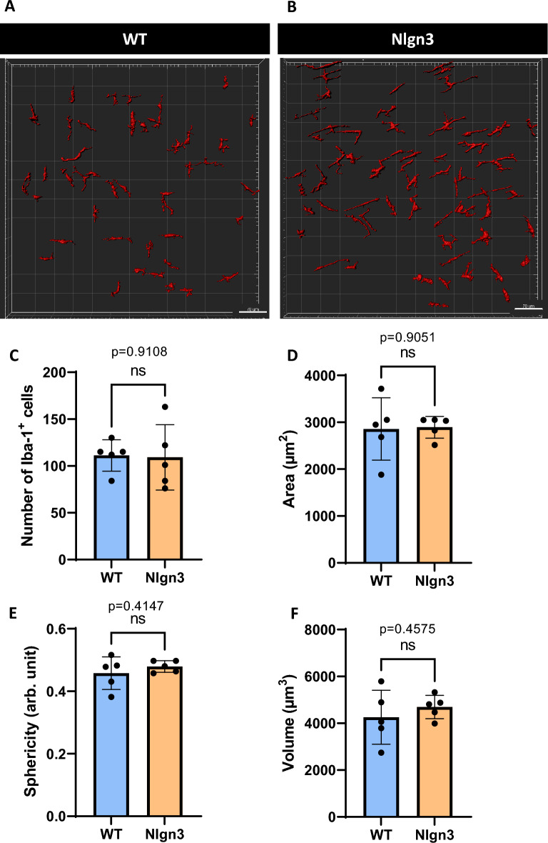
Figure 9.
No morphological difference was observed in Iba-1+ immunopositive cells in the myenteric plexus of Nlgn3R451C cecum compared to WT littermates. 3D reconstruction and analysis of Iba-1+ cells in WT (A) and Nlgn3 (B) myenteric plexus. No significant morphological differences were observed with the parametrized batches of Iba-1+ cells for (C), area (D), sphericity (E), and volume (F). Statistical comparisons were conducted using Student’s unpaired t-test. Individual data and mean ± SD were plotted for n = 5 mice in each group. ns = p > 0.05. WT = wildtype; Nlgn3 = Nlgn3R451C.

