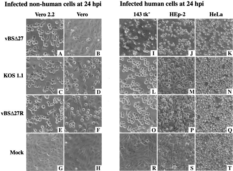
FIG. 1.
Morphologies of infected nonhuman (A to H) Vero 2.2 and Vero cells and human (I to T) 143tk−, HEp-2, and HeLa cell lines. Cells infected with vBSΔ27 (A, B, and I to K), KOS1.1 (C, D, and L to N), or vBSΔ27R (E, F, and O to Q) and mock-infected cells (G, H, and R to T) were observed at 24 h p.i. by phase-contrast light microscopy (magnification, ×20) as described in Materials and Methods.

