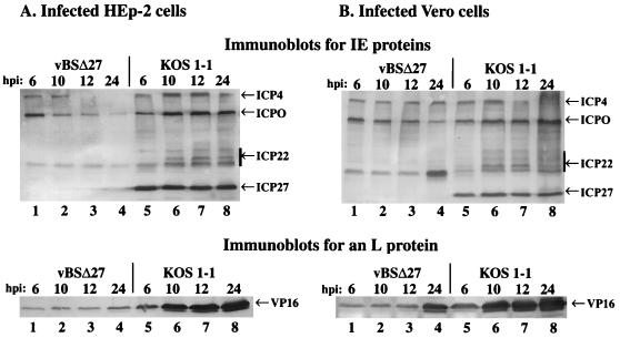
FIG. 3.
Accumulation of IE and L proteins in infected cells at various infection times. Total cell extracts (50 μg) prepared at 6, 10, 12, and 24 h p.i. from vBSΔ27- and KOS1.1-infected HEp-2 (A) and Vero (B) cells were used for immunoblot analyses with the polyclonal anti-ICP22 antibody (RGST22), monoclonal anti-ICP4 (1114), anti-ICP27 (1113), anti-ICP0 (1112) antibodies (IE proteins), and the monoclonal anti-VP16 antibody (L protein). IE and L protein locations are shown in the right margins (arrows), and the bars indicate the multiple electrophoretic forms of ICP22.

