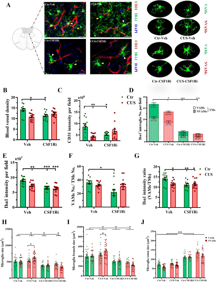
Fig. 5.
CUS and CSF1Ri reduced CD31+-blood vessels and differentially affected VAMs and NVAMs in mice. (A) Representative staining of CD31 and IBA1 in the PFC (scale bar = 10 µm) with enlarged VAMs and NVAMs (indicated by white and yellow arrowheads, respectively) are shown. (B) Blood vessel density (e.g., vessel area/total area*100%) and (C) total CD31 intensity. (D) TMs-(including VAMs and NVAMs)-No. and (E) total IBA1 intensity. (F) Ratio of VAMs-No./TMs-No. and (G) ratio of IBA1 intensity in VAMs/TMs. (H) Microglia cell size, (I) branch size and (J) cell soma size (n = 3 mice/12 sections/40 ~ 200 cells per group). Ctr: control; CUS: chronic unpredictable stress; CSF1Ri: CSF1R inhibitor; No.: number; TM: total microglia/macrophages; Veh: vehicle; VAMs: vessel-associated microglia/macrophages; NVAMs: non-vessel-associated microglia/macrophages. Data presented as mean ± SEM; */**/*** p < 0.05/0.01/0.001. Two-way ANOVA with Bonferroni’s correction. See also Additional file 2: Fig. S3 & S4

