Figure 1.
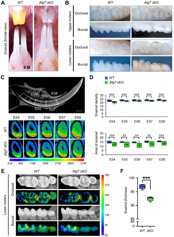
Gross appearance of the teeth of wild-type and Atg7 mutant mice. (A) Incisors of wild-type (WT) and Atg7 conditional knockout (cKO) mice at 6 mo (6 M) of age. (B) Molars of WT and Atg7 cKO mice at 6 mo of age. (C) The region containing enamel in the lower incisor was divided into eight 1-mm-long cross sections (ES1–ES8) from near the apical loop to the gingival margin. Upper panel: a representation of a model WT tooth with sagittal micro–computed tomography (CT) sections (ES4–ES8) of the lower incisor at 6 mo of age. Lower panels: transverse micro-CT sections, taken at the indicated time points, of lower incisors from WT and Atg7 cKO mice. Scale bar: 1 mm. Color bar, density in Hounsfield units, range 516 to 3,100. (D) Quantification of mineral density and area of the enamel in incisors from WT and Atg7 cKO mice at 6 mo of age. **P < 0.01. ***P < 0.001. n = 6 per group. (E) Micro-CT images of molars from WT and Atg7 cKO mice at 6 mo of age. Color bar, density in Hounsfield units, range 60 to 350. (F) Quantification of enamel thickness in molars from WT and Atg7 cKO mice at 6 mo of age. ***P < 0.001. n = 6 per group.

