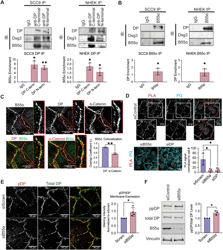
Figure 2.
DP is found in complex with the PP2A regulatory subunit B55⍺ in SCC9 cells. (A) Immunoprecipitation of endogenous DP using antibodies targeting DP’s C-terminus or N-terminus and blotting back for Dsg3 or B55⍺. SCC9s (left) and NHEKs (right) were grown for 2 days in high-calcium media (HCM). (B) Immunoprecipitation of endogenous B55⍺ and blotting back for DP or Dsg3. SCC9s (left) and NHEKs (right) were grown for 2 days in high-calcium media (HCM). (C) SCC9 immunofluorescence co-stained for B55⍺, DP, and ⍺-Catenin. Overlayed images are shown below. Colocalization analysis as determined by an object-based colocalization analysis tool represented as overlap coefficient measurements. (D) Proximity ligation analysis performed on SCC9 cell transfected with siRNA targeting either a Scramble control, B55α, or DP. A fluorescence-based PLA signal was measured on fixed coverslips incubated with B55α and DP targeting antibodies. (E) Immunofluorescence staining of S2849 phosphorylated DP (pDP) in SCC9 cells transfected with siRNA targeting the B55α subunit. Staining intensity from was quantified at the membrane using PG stain as a mask. (F) Amount of the dual S2845/S2849 phosphorylated DP (ppDP) were analyzed in total SCC9 cells transfected with siRNA targeting the B55α subunit. Statistical analyses were performed using a One-way ANOVA with multiple comparisons (A,D) or a student t-test (B–C,E–F). * < 0.05; ** < 0.01; *** < 0.001.

