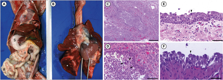
Fig. 3. Necropsy and histologic findings. (A) Gross findings of the abdominal organs. All internal organs show complete left-to-right transposition of the thoracic and abdominal organs. (B) Gross findings of the lungs. Severe diffuse collapse, congestion, and hemorrhages of lung lobes. (C) Severe diffuse purulent bronchopneumonia of the lung. (D) Large eosinophilic and basophilic intranuclear inclusion bodies are observed in the detached epithelial cells in the bronchiolar lumen (arrow heads). (E) Severe diffuse deciliation, mild diffuse desquamation of the epithelial cells, and mild diffuse lymphoid cell infiltrations in the submucosa. Note: basophilic intranuclear inclusion body (arrowhead). (F) Transitional epithelial cells in the urinary bladder contain small eosinophilic intracytoplasmic inclusion bodies. Staining: hematoxylin and eosin. Scale bar: (C) 200 μm, (D) 50 μm, (E, F) 25 μm.
L, left; R, right; S, stomach; H, heart.

