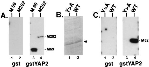
FIG. 5.
Far-Western analysis of GST-rabies virus M fusion proteins. (A) Duplicate nitrocellulose filters with gstRabM69 (M 69; lanes 1 and 3) and gstRabM202 (containing the full-length M protein M202; lanes 2 and 4) were probed with either GST alone (lanes 1 and 2) or gstYAPWW2 (lanes 3 and 4). (B) Coomassie brilliant blue stain of bacterial cell extracts expressing approximately 1.0 μg of gstRabM52Y-A (Y>A; lane 1) or gstRabM52WT (WT [wild type]; lane 2) indicated by the arrowhead. (C) Identical amounts (1.0 μg/lane) of gstRabM52Y-A and gstRabM52WT to those seen in panel B were immobilized onto duplicate nitrocellulose filters and probed with either GST alone (lanes 1 and 2) or gstYAPWW2 (lanes 3 and 4).

