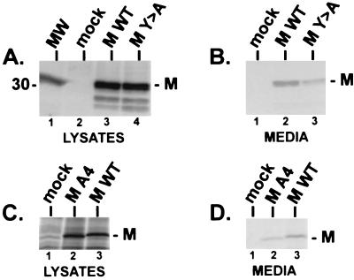
FIG. 7.
VSV M budding assay. (A) Radiolabeled lysates from CV-1 cells receiving no DNA (mock, lane 2), T7VSVMWT DNA (MWT, lane 3), and T7VSVMY-A DNA (MY>A, lane 4) were immunoprecipitated with polyclonal antiserum against the M protein of VSV and fractioned by SDS-PAGE. The position of the M protein of VSV is indicated. MW, 14C-labeled protein standards. (B) Radiolabeled proteins released into the media covering cells transfected with no DNA (mock, lane 1), T7VSVMWT DNA (lane 2), and T7VSVMY-A DNA (lane 3) were immunoprecipitated with polyclonal antiserum against the M protein of VSV and fractionated by SDS-PAGE. The position of the M protein of VSV is indicated. (C) Radiolabeled lysates from CV-1 cells receiving no DNA (mock, lane 1), T7VSVMA4 DNA (lane 2), and T7VSVMWT DNA (lane 3) were immunoprecipitated with polyclonal antiserum raised against VSV virions (ATCC) and fractionated by SDS-PAGE. (D) Radiolabeled proteins released into the media covering cells transfected with no DNA (mock, lane 1), T7VSVMA4 (lane 2), and T7VSVMWT (lane 3) were immunoprecipitated with polyclonal antiserum raised against VSV virions (ATCC) and fractionated by SDS-PAGE.

