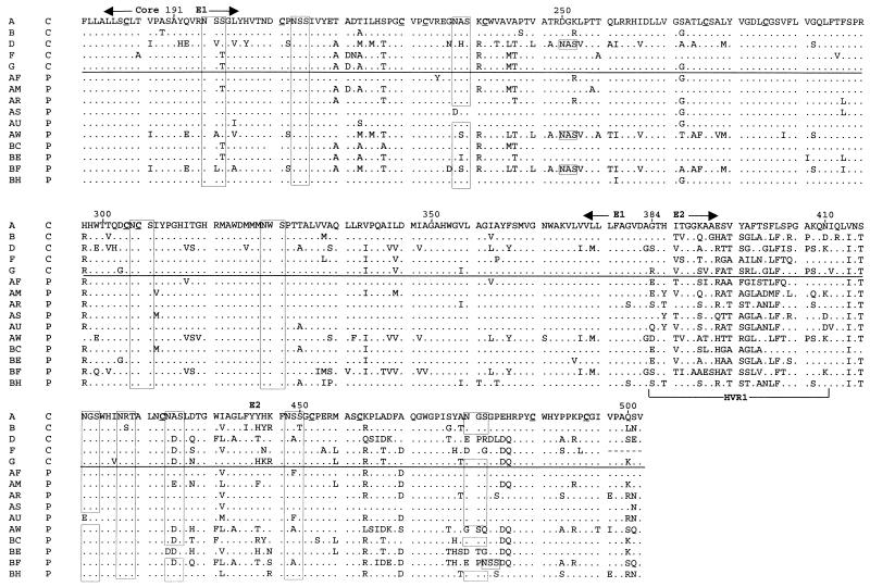
FIG. 2.
Alignment of inferred amino acid sequences for the majority sequences from each subject. In the first column, an alphabetical label is given for each subject, while in the second column, C indicates clearance of viremia and P indicates persistence. Periods indicate identity to the amino acid at that position in the first sequence. Positions of the Core and E1 and E2 regions are indicated above the alignment, whereas that of HVR1 is indicated below the alignment at the N terminus of E2. Boxes indicate predicted N-linked glycosylation sites. Cysteine residues in the first sequence are underlined.

