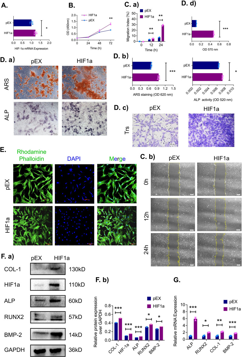
Fig. 2.
HIF-1α overexpression in ADSCs significantly increases cell proliferation and osteogenic capacity in vitro. A The mRNA expression of HIF-1α in ADSCs after transfection with a HIF-1α overexpression plasmid and an empty plasmid (pEX) was evaluated by qRT‒PCR (n = 3). B The proliferation of transfected ADSCs was evaluated by a CCK-8 assay (n = 3). C a, b The scratch assays at 0, 12 and 24 h (n = 3). D a, b ARS and BCIP/NBT staining after osteogenic induction. D c, d Representative images and quantitative analysis of Transwell assays after 24 h (n = 3, scale bar = 100 μm). E Representative images of ADSC focal adhesion plaques using immunofluorescence staining (F-actin in green and nuclei in blue, scale bar = 100 μm). F a, b Protein levels and (G) mRNA and of the osteogenic marker genes (ALP, RUNX2, COL1, and BMP2). *p < 0.05; **p < 0.01; ***p < 0.001

