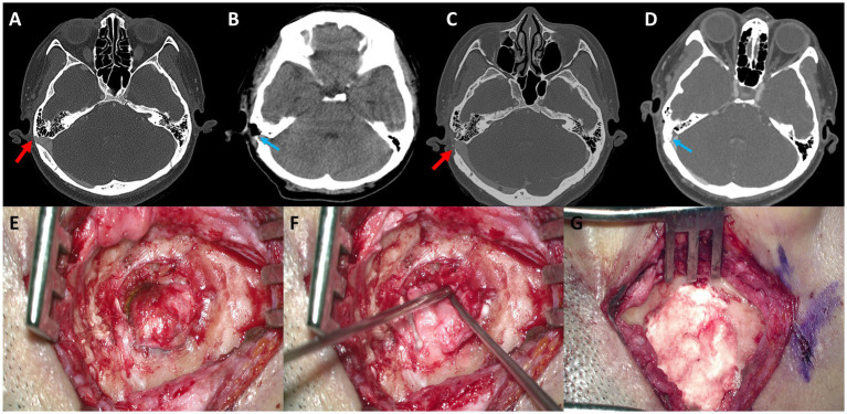
Figure 4.
(A) Preoperative axial temporal bone computed tomography (TBCT) image of Subject 4 shows a huge diverticulum protruding through the mastoid air cells and cortical mastoid bone to the level of the subcutaneous soft tissue layer (arrow). (B) Postoperative axial TBCT image shows successful reduction of the diverticulum through transmastoid resurfacing with bone wax (arrow) and fibrin glue. (C) Follow-up TBCT axial image shows a huge diverticulum protruding through the mastoid air cells and cortical mastoid bone to the level of the subcutaneous soft tissue layer (arrow). (D) Again, the diverticulum is successfully reduced by transmastoid reshaping with harvested autologous cortical bone chips and bone cement (arrow). (E) The diverticulum was exposed to the mastoid cortex again. (F,G) Thus, revision reshaping of the sigmoid sinus diverticulum was performed with harvested autologous cortical bone chips and bone cement to reconstruct a secure sinus wall over the diverticulum.

