
Figure 6.
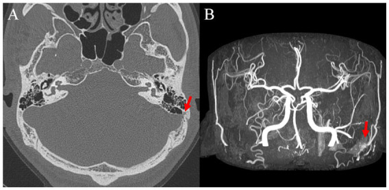
(A) Preoperative axial temporal bone computed tomography image showing left sigmoid sinus dehiscence and (B) postoperative brain magnetic resonance image with angiography image showing left dural arteriovenous fistula.
Official websites use .gov
A
.gov website belongs to an official
government organization in the United States.
Secure .gov websites use HTTPS
A lock (
) or https:// means you've safely
connected to the .gov website. Share sensitive
information only on official, secure websites.
(A) Preoperative axial temporal bone computed tomography image showing left sigmoid sinus dehiscence and (B) postoperative brain magnetic resonance image with angiography image showing left dural arteriovenous fistula.