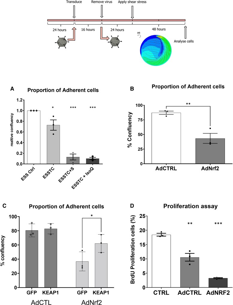
Figure 2.
Evidence for a role for Nrf2 in endothelial detachment. (A) Quantification of cell number in elevated laminar shear stress control (ESS), with the addition of TNFα and CSE [mean ± SD, one-way ANOVA, elevated flow + TNFα + CSE (ESSTC), 30% reduction vs. ESS, P < 0.05, n = 3], and Nrf2 activator sulforaphane (ESSTC-S, 2.5 μM, 5-fold reduced adhesion vs. ESSTC, P < 0.05, n = 3) or isoliquiritigenin (ESS-IsoQ, 10 μM, 9-fold reduced adhesion vs. ESSTC, P < 0.05, n = 3). (B) Adenoviral overexpression of Nrf2 (200 pfu/cell and 200 pfu/cell AdCTRL combined to match later experiments) promotes 50% of cell detachment compared with AdCTRL (400 pfu/cell) (****P < 0.0001, n = 3) using the experimental design illustrated above. (C) Transduction with lentiviral control (GFP) or lentiKEAP1 prior to adenoviral overexpression of Nrf2 (as C) resulted in a significant reduction of Nrf2-dependent detachment (*P < 0.05, n = 3). (D) BrdU proliferation assay in HCAECs treated with adenoviral overexpression of wild-type Nrf2, % BrdU positive cells, mean and SEM obtained from n = 6, *P < 0.05 and **P < 0.01, compared with control.

