Figure 1:

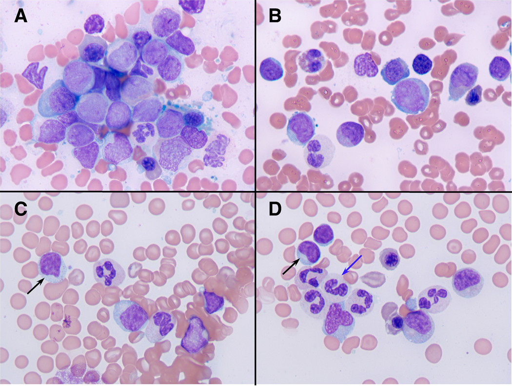
Bone marrow morphology before (A, B) and after cord blood stem cell transplantation (C, D). Fig. 1 A and B depict bone marrow aspirate smears at the initial presentation with myeloblasts in a background of dysplastic erythroid and myeloid elements (Wright-Giemsa stains, photographed at 1000x). Fig. 1 C and D display photomicrographs of bone marrow aspirate smears after CB-SCT that are consistent with a complete remission and that depict scattered large granular lymphocytes (black arrows) in a background of mildly dysplastic myeloid cells, some with hypogranular cytoplasm (blue arrow).
