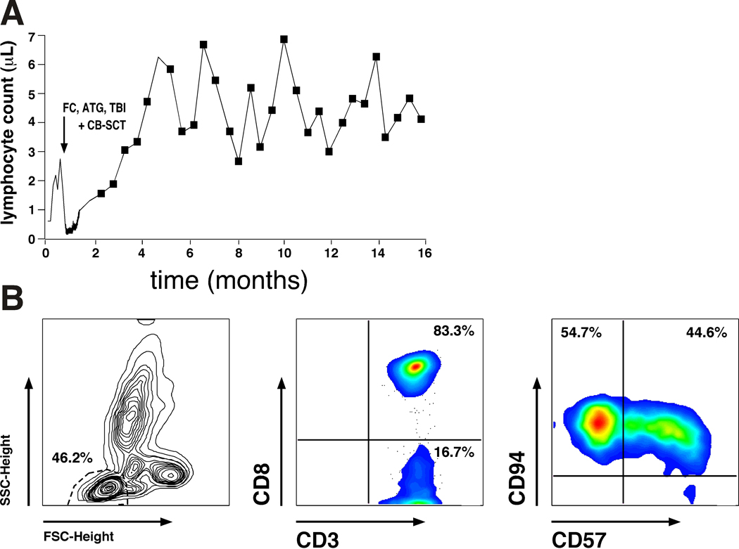
Figure 2:
Expansion of autologous, CD8+ T cells after CB-SCT. Fig. 2A depicts the kinetics of the lymphocytosis after non-myeloablative conditioning with FC, ATG and TBI, and subsequent CB-SCT (indicated by the arrow). Fig. 2B illustrates the immunophenotype of the expanded T cells. Displayed are the forward (FSC) and sideward scatter (SSC) characteristics with gating on the lymphocyte population (left hand box), and staining with anti-CD3 and anti-CD8 mAbs, revealing that 83.3% of the lymphocytes were CD8 positive (center box). Staining with anti-CD57 and anti-CD94 mAbs (right hand box) also revealed that these CD8+ T cells displayed a phenotype consistent with large granular lymphocytes (LGL), which is in keeping with the morphology of these activated lymphocytes (Fig. 1C, D).

