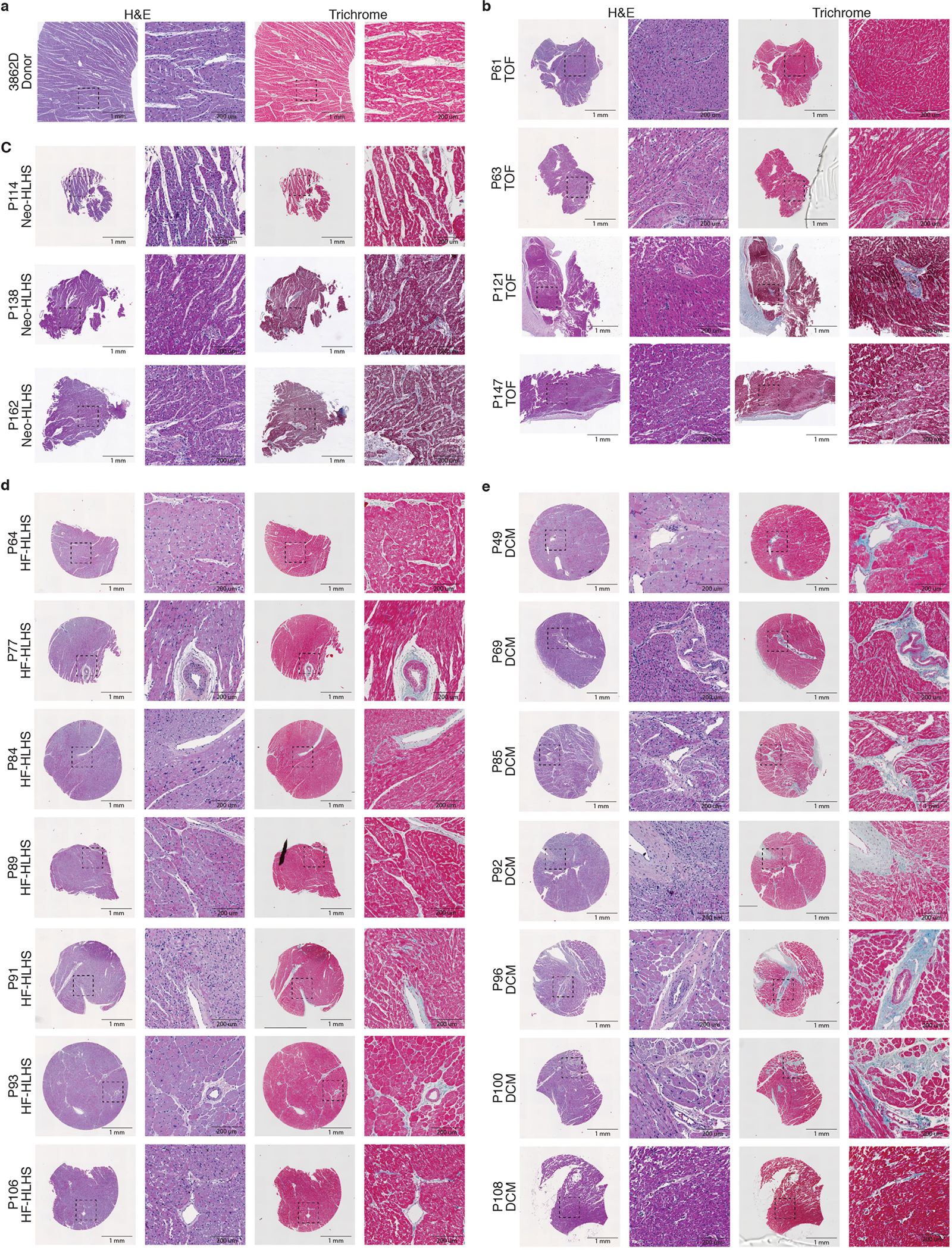
Extended Data Fig. 6 |. Additional tissue histology.

a–e, H&E and trichrome staining of additional myocardial samples from donor (a), TOF (b), Neo-HLHS (c), HF-HLHS (d), and DCM (e) patients. Left image is a 2-mm core, and the dashed box outlines the highlighted perivascular region at high magnification in the right image.
